LATERAL ELBOW PROJECTION
Lateral projection protocol for elbow joint
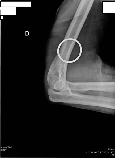
Important Note About Medical Devices
The visible rings in the radiograph correspond to a sling or arm sling
The immobilization device should not be removed until the full extent of the injury is observed in the radiographs.
This precaution is essential to avoid aggravating injuries during the radiological evaluation process.
Exposure Factors
Low exposure: Parameters optimized for lateral visualization of elbow joint
Visible Anatomical Structures
Should be clearly observed:
- Elbow joint in true lateral view
- Distal part of humerus (superimposed condyles)
- Proximal part of ulna (olecranon in profile)
- Proximal part of radius (radial head)
- Humeroulnar joint space in profile
- Proximal radioulnar relationship
- Olecranon fossa of humerus
Plate Size and Division
Standard plate for elbow
To perform AP and lateral projections on a single plate
Divided plate: The second portion of the plate is used to complete the study with lateral view
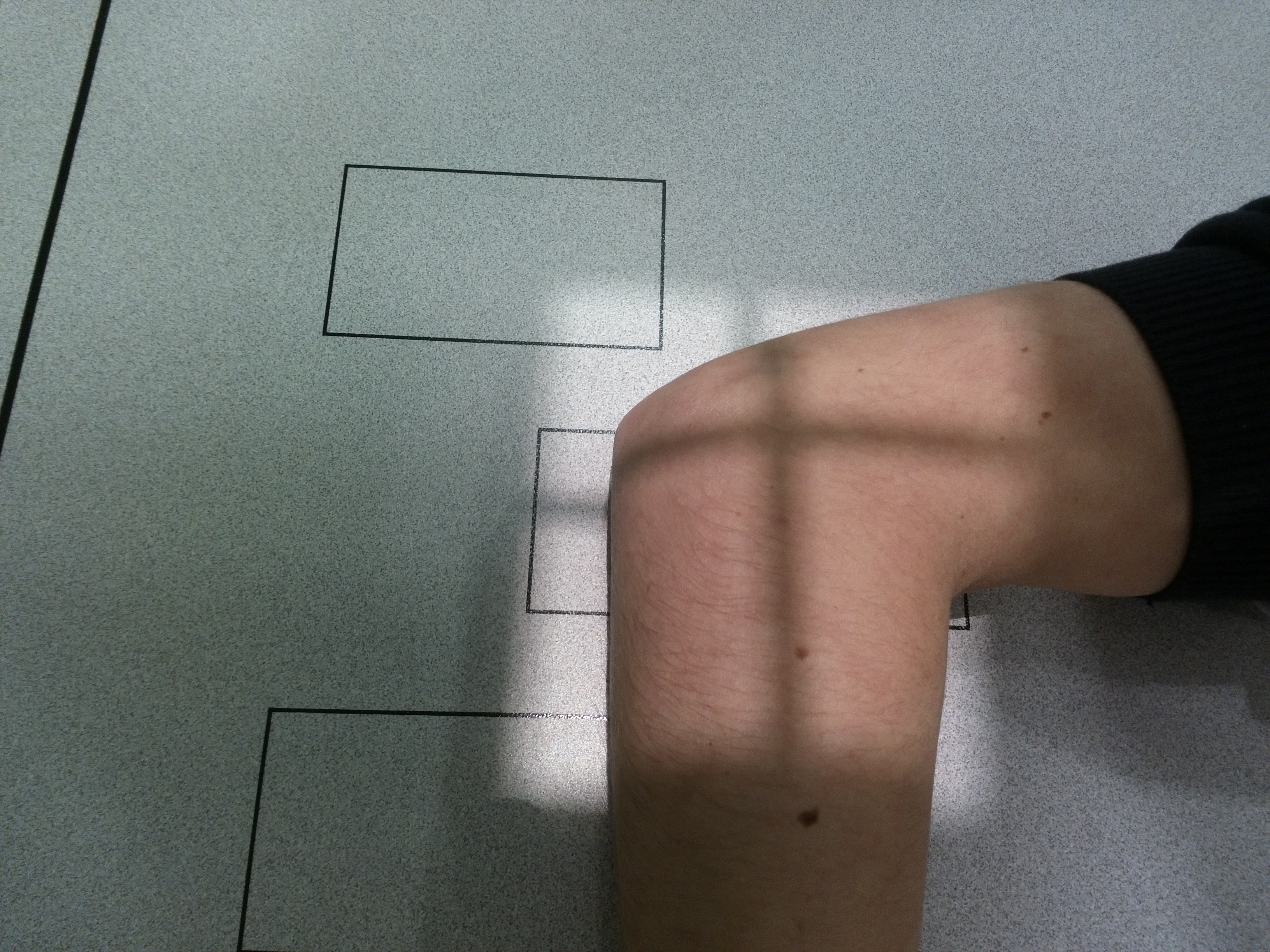
Patient Positioning
Critical Alignment Point
To obtain a true lateral projection of elbow, humeral epicondyles (medial and lateral) must be perfectly aligned in perpendicular position to chassis plane.
This ensures correct superimposition of humeral condyles in radiographic image.
Central Ray Point
Direction: Vertical and perpendicular to joint center
Location: Lateral humeral epicondyle (radial condyle)
Target: Humeroradial joint space
Specific Hand and Elbow Position
Hand Position
Hand in lateral position
Thumb up
Neutral forearm pronation
Elbow Flexion
Exact 90° flexion
Support on medial surface
Arm and forearm in same plane
Considerations for Injured Patients
Limited Flexion
If 90° flexion not possible:
• Flex to maximum tolerated
• Document flexion degree
• Consider alternative projection
Immobilization Devices
Keep sling during study
Do not remove until complete evaluation
Note its presence in report
Patient Instructions
"Do not move during exposure"
Maintain position without movement during radiographic exposure
Special attention to maintain elbow flexed 90° and hand with thumb up
Optimal Image Characteristics
True lateral view
Humeral condyles superimposed
Olecranon in profile
Process clearly visible
Joint spaces
Humeroulnar and humeroradial visible
Adequate field
Distal humerus to proximal ulna/radius
Common Technical Challenges
Frequent problems in lateral elbow projection:
- Arm rotation causing oblique view instead of true lateral
- Incorrect flexion of elbow (not exact 90°)
- Poor epicondylar alignment (not perpendicular to chassis)
- Incorrect hand position (not lateral with thumb up)
- Incorrect support (not on medial surface)
- Incomplete superimposition of humeral condyles
- Movement during exposure due to poor stabilization
Solution: Verify humeral epicondyles are perpendicular to chassis and maintain exact 90° flexion with medial support